![Intraoral camera in [city], [st]: A closer look at your smile](/images/internalbannerbg.webp)
Intraoral camera in [city], [st]: A closer look at your smile
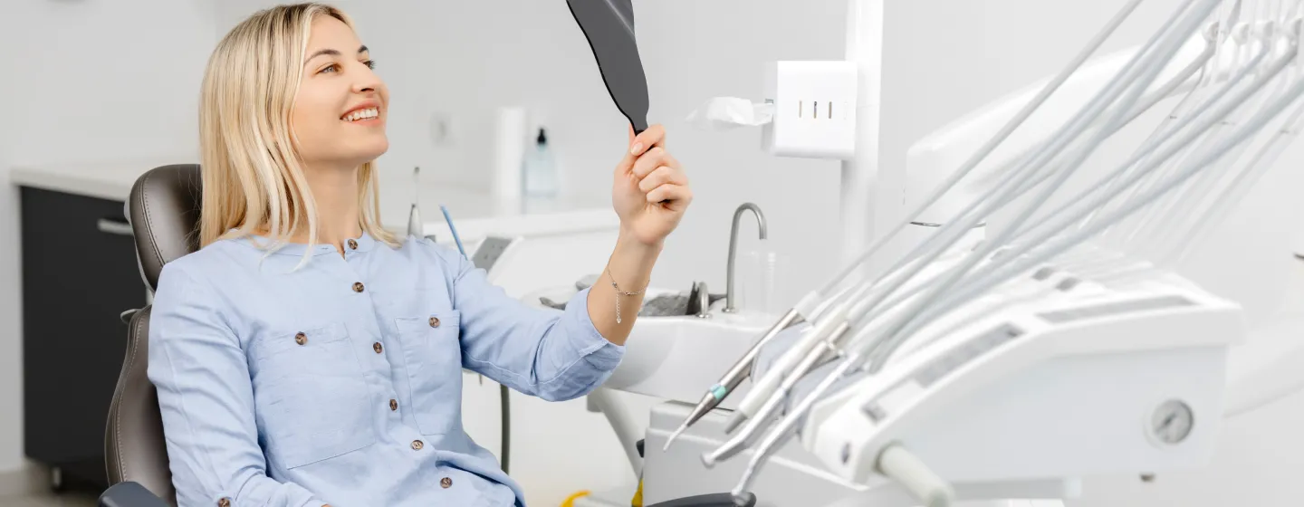
At Summit View Dental & Wellness Center in Millcreek, UT, an intraoral camera gives a high-resolution view of hard-to-see areas of your mouth. This small device helps patients see what [dr_type2] sees and supports accurate diagnoses and treatment planning. If you have questions about the intraoral camera in Millcreek, UT or want to review images from a recent visit, call 801-561-0061.
Intraoral camera explained
An intraoral camera is a pen-sized digital camera that captures detailed photos and short videos of your teeth and gums. The images appear on a chairside monitor in real time, allowing clear visualization of cracks, chips, decay, worn fillings, gum inflammation, and other conditions. Unlike dental X-rays, which use radiation to show structures beneath the surface, this digital dental imaging tool uses light to show what is visible within the mouth.
Patients often search for what is an intraoral camera and how does an intraoral camera work. The camera sits inside a protective sheath and rests gently on or near the tooth. With magnification and LED lighting, it reveals fine details that can be missed by the naked eye. The result is a shared view that improves chairside patient education and documentation.
How intraoral images can help you
- Early cavity detection is possible because magnified images highlight small areas of demineralization or staining.
- Cracks and fractures are easier to see, helping differentiate between surface craze lines and structural fractures.
- Gum health can be monitored by photographing inflammation, recession, and plaque buildup over time.
- Treatment discussions are clearer when you can see the area being treated on a screen during your consultation.
- Progress tracking is simple because before-and-after images document results after fillings, crowns, or cosmetic care.
- Insurance documentation may be supported by clear photographs of conditions requiring treatment.
The imaging process
The process is quick and comfortable. Here is a step-by-step look at what typically happens during a visit.
- The camera is covered with a new, disposable barrier to maintain strict infection control.
- Your clinician positions the camera to capture high-resolution images of specific teeth or gum areas.
- The images display instantly on a chairside monitor so you can follow along in real time.
- Photos are saved to your secure digital chart for future comparison and treatment planning.
- The disposable cover is removed and discarded, and the device is disinfected according to clinical protocols.
What to expect
Most patients find the intraoral camera comfortable because it is slim and used briefly in each area. The process usually adds only a few minutes to your appointment. There is no radiation exposure because the device uses light, not X-rays. The camera complements dental X-rays rather than replacing them, so both tools may be recommended depending on your needs.
You do not need special preparation. Tooth surfaces may be dried with air for a clearer image, and your lips or cheeks may be gently retracted for visibility. If you have a sensitive gag reflex, you can ask for short breaks while images are taken. Images can be reviewed together on the screen, making it easier to discuss the benefits of an intraoral camera and your next steps.
Like any tool, the camera has limitations. It cannot show hidden structures such as the nerve or bone level, which is why X-rays and other tests are still important. Still, the combination of visual photos and radiographs provides a more complete picture for accurate diagnosis and follow-up care.
FAQs
Contact Us
Contact Us
Schedule Your Visit
Ready to experience exceptional dental care? Contact our team today to schedule your appointment. We're here to answer your questions and help you achieve optimal oral health.